38 microscope images with labels
Microscope Labels Photos - Free & Royalty-Free Stock Photos from Dreamstime Download all free or royalty-free photos and images. Use them in commercial designs under lifetime, perpetual & worldwide rights. Dreamstime is the world`s largest stock photography community. Label Microscope Diagram - EnchantedLearning.com Using the terms listed below, label the microscope diagram. arm - this attaches the eyepiece and body tube to the base. base - this supports the microscope. body tube - the tube that supports the eyepiece. coarse focus adjustment - a knob that makes large adjustments to the focus. diaphragm - an adjustable opening under the stage, allowing ...
label the microscope worksheet Microscope Worksheet Label. 16 Images about Microscope Worksheet Label : Parts of a Microscope Labeling Worksheet - Science by TechCheck Lessons, Glossary of terms used in microscopy - Quekett Microscopical Club and also 32 Label The Microscope Worksheet - Labels For You.

Microscope images with labels
Microscope Images Labeled | Virtual Anatomy Lab VAL - ncccval Body cavities, planes, and regions. Body Images Labeled. Body Images Unlabeled. Histology. Epithelium Images Labeled. Epithelium Images Unlabeled. Connective Tissue Images Labeled. Connective Tissue Images Unlabeled. Microscope. Parts of a microscope with functions and labeled diagram - Microbe Notes Parts of a microscope with functions and labeled diagram September 17, 2022 by Faith Mokobi Having been constructed in the 16th Century, Microscopes have revolutionalized science with their ability to magnify small objects such as microbial cells, producing images with definitive structures that are identifiable and characterizable. Amazon.com: microscope slide labels Microscope Slide Label SLS-15, Standard, 1000/PK. $33.00 $ 33. 00. Get it Wed, Oct 19 - Mon, Oct 24. $8.00 shipping. Only 10 left in stock - order soon. Small Business. Small Business. Shop products from small business brands sold in Amazon's store. Discover more about the small businesses partnering with Amazon and Amazon's commitment to ...
Microscope images with labels. Label the microscope — Science Learning Hub All microscopes share features in common. In this interactive, you can label the different parts of a microscope. Use this with the Microscope parts activity to help students identify and label the main parts of a microscope and then describe their functions. Drag and drop the text labels onto the microscope diagram. Microscope Images at Various Magnifications | Microscope World Resources The compound microscope typically has three or four magnifications - 40x, 100x, 400x, and sometimes 1000x. At 40x magnification you will be able to see 5mm. At 100x magnification you will be able to see 2mm. At 400x magnification you will be able to see 0.45mm, or 450 microns. At 1000x magnification you will be able to see 0.180mm, or 180 microns. Microscope Labeling Game - PurposeGames.com About this Quiz. This is an online quiz called Microscope Labeling Game. There is a printable worksheet available for download here so you can take the quiz with pen and paper. This quiz has tags. Click on the tags below to find other quizzes on the same subject. Science. Microscope Types (with labeled diagrams) and Functions The working principle of a simple microscope is that when a lens is held close to the eye, a virtual, magnified and erect image of a specimen is formed at the least possible distance from which a human eye can discern objects clearly. Simple microscope labeled diagram Simple microscope functions It is used in industrial applications like:
Microscope Parts, Function, & Labeled Diagram - slidingmotion Microscope parts labeled diagram gives us all the information about its parts and their position in the microscope. Microscope Parts Labeled Diagram The principle of the Microscope gives you an exact reason to use it. It works on the 3 principles. Magnification Resolving Power Numerical Aperture. Parts of Microscope Head Base Arm Eyepiece Lens Skin Images Labeled | Virtual Anatomy Lab VAL - ncccval Body cavities, planes, and regions. Body Images Labeled. Body Images Unlabeled. Histology. Epithelium Images Labeled. Epithelium Images Unlabeled. Connective Tissue Images Labeled. Connective Tissue Images Unlabeled. Microscope. Microscope Labeling - The Biology Corner The google slides shown below have the same microscope image with the labels for students to copy. I often spend the first day walking students through the steps and having them look at a single slide as we do the steps. Students are often very enthusiastic about using microscopes and will try to start with the high power objective. Microscope Photos and Premium High Res Pictures - Getty Images Browse 46,685 microscope stock photos and images available, or search for magnifying glass or microscope isolated to find more great stock photos and pictures. magnifying glass. microscope isolated. science.
Parts of the Microscope with Labeling (also Free Printouts) Microscopes are specially created to magnify the image of the subject being studied. This exercise is created to be used in homes and schools. the microscope layout, including the blank and answered versions are available as pdf downloads. Click to Download : Label the Parts of the Microscope (A4) PDF print version. Explanation and Labelled Images - New York Microscope Company Another use of fluorescence imaging is Fluorescence Speckle Microscopy. It is a technology that uses fluorescence labeled macromolecular assemblies such as cytoskeletal protein to study movement and turnover rates. Fluorescence microscopy staining also is helpful in the field of mineralogical applications. It is routinely used for the study of ... Compound Microscope Parts - Labeled Diagram and their Functions The eyepiece (or ocular lens) is the lens part at the top of a microscope that the viewer looks through. The standard eyepiece has a magnification of 10x. You may exchange with an optional eyepiece ranging from 5x - 30x. [In this figure] The structure inside an eyepiece. The current design of the eyepiece is no longer a single convex lens. Microscope Labeled Pictures, Images and Stock Photos Browse 49 microscope labeled stock photos and images available, or start a new search to explore more stock photos and images. Newest results Fluorescent Imaging immunofluorescence of cancer cells growing... Microscope diagram vector illustration. Labeled zoom instrument... Microscope diagram vector illustration.
Label a Microscope Worksheet
400+ Free Microscope & Bacteria Images - Pixabay 413 Free images of Microscope Related Images: bacteria science laboratory research scientist biology lab chemistry microbiology Find your perfect microscope image. Free pictures to download and use in your next project. Next page ›
Label microscope - Teaching resources
Microscope Pictures, Images and Stock Photos Browse 201,468 microscope stock photos and images available, or search for magnifying glass or microscope isolated to find more great stock photos and pictures. Microscope icon. Medical diagnostics, laboratory, research, education, study. Vector illustration. Equipment for medical research, laboratories.

Lable the microscope worksheet
Labeling the Parts of the Microscope | Microscope World Resources Labeling the Parts of the Microscope This activity has been designed for use in homes and schools. Each microscope layout (both blank and the version with answers) are available as PDF downloads. You can view a more in-depth review of each part of the microscope here. Download the Label the Parts of the Microscope PDF printable version here.

Parts of a Microscope - SmartSchool Systems
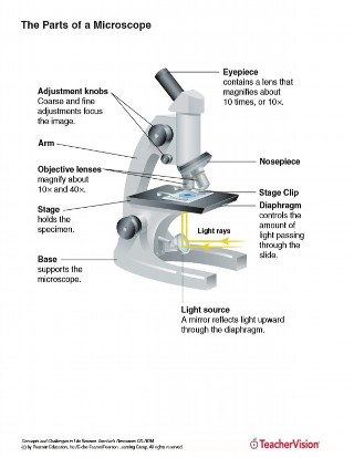
Microscope Parts and Functions Body tube (Head): The body tube connects the eyepiece to the objective lenses. Arm: The arm connects the body tube to the base of the microscope. Coarse adjustment: Brings the specimen into general focus. Fine adjustment: Fine tunes the focus and increases the detail of the specimen. Nosepiece: A rotating turret that houses the objective lenses.

Microscope Labels Flashcards | Quizlet
Amazon.com: microscope slide labels Microscope Slide Label SLS-15, Standard, 1000/PK. $33.00 $ 33. 00. Get it Wed, Oct 19 - Mon, Oct 24. $8.00 shipping. Only 10 left in stock - order soon. Small Business. Small Business. Shop products from small business brands sold in Amazon's store. Discover more about the small businesses partnering with Amazon and Amazon's commitment to ...

Solved Microscope parts/labeling 9 Label the image of a ...
Parts of a microscope with functions and labeled diagram - Microbe Notes Parts of a microscope with functions and labeled diagram September 17, 2022 by Faith Mokobi Having been constructed in the 16th Century, Microscopes have revolutionalized science with their ability to magnify small objects such as microbial cells, producing images with definitive structures that are identifiable and characterizable.

Virtually Labeling a Microscope by Grace Voit | Teachers Pay ...
Microscope Images Labeled | Virtual Anatomy Lab VAL - ncccval Body cavities, planes, and regions. Body Images Labeled. Body Images Unlabeled. Histology. Epithelium Images Labeled. Epithelium Images Unlabeled. Connective Tissue Images Labeled. Connective Tissue Images Unlabeled. Microscope.
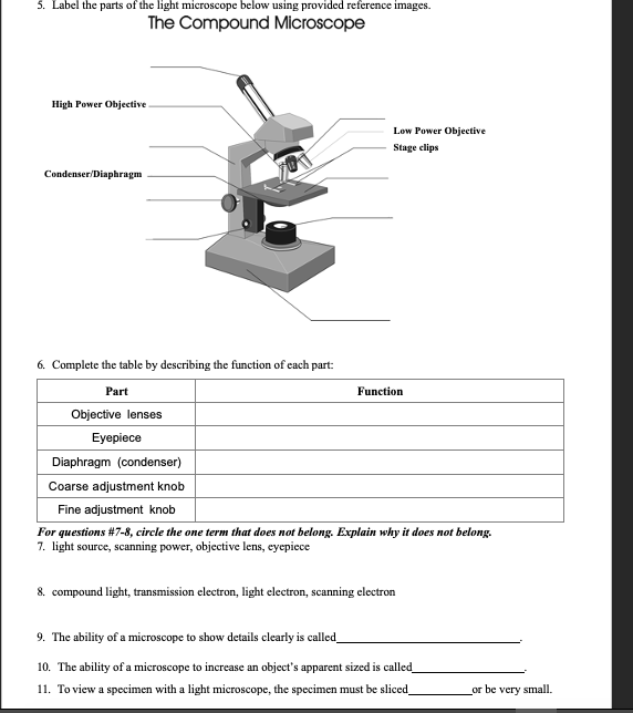
Solved 5. Label the parts of the light microscope below ...

Microscope With Labels clip art | Microscope parts ...

Microscope Labeling Activity - SMART Board Activity - Interactive Review

Label The Microscope Diagram - Robot PNG Image | Transparent ...
Label a microscope - Teaching resources

Below is a photo of a compound light microscope with labels ...
4,874 Microscope Labeled Images, Stock Photos & Vectors ...

Compound Microscope Parts – Labeled Diagram and their ...

Meiji MT6500 Series PCM NIOSH 7400 Asbestos Microscope
Label microscope - Teaching resources
Microscope slide Vector Art Stock Images | Depositphotos

Microscope Components - Science Quiz
microscope with labels - Openclipart
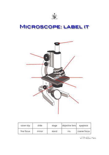
Microscope: label it | Teaching Resources
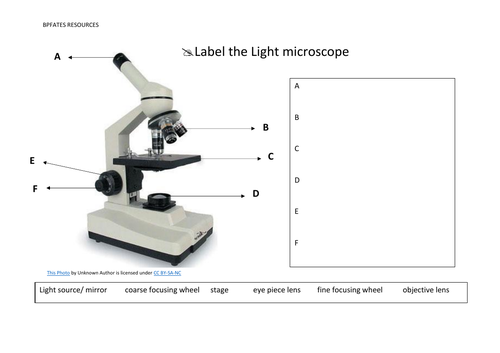
Label the light microscope | Teaching Resources

Compound Microscope Parts, Diagram Definition, Application ...
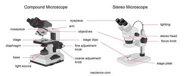
Parts of Stereo Microscope (Dissecting microscope) – labeled ...
Getting Started - Virtual Fluorescent Microscope - Wartburg ...

Parts of Stereo Microscope (Dissecting microscope) – labeled ...

This is a common compound microscope. Label its parts from A ...
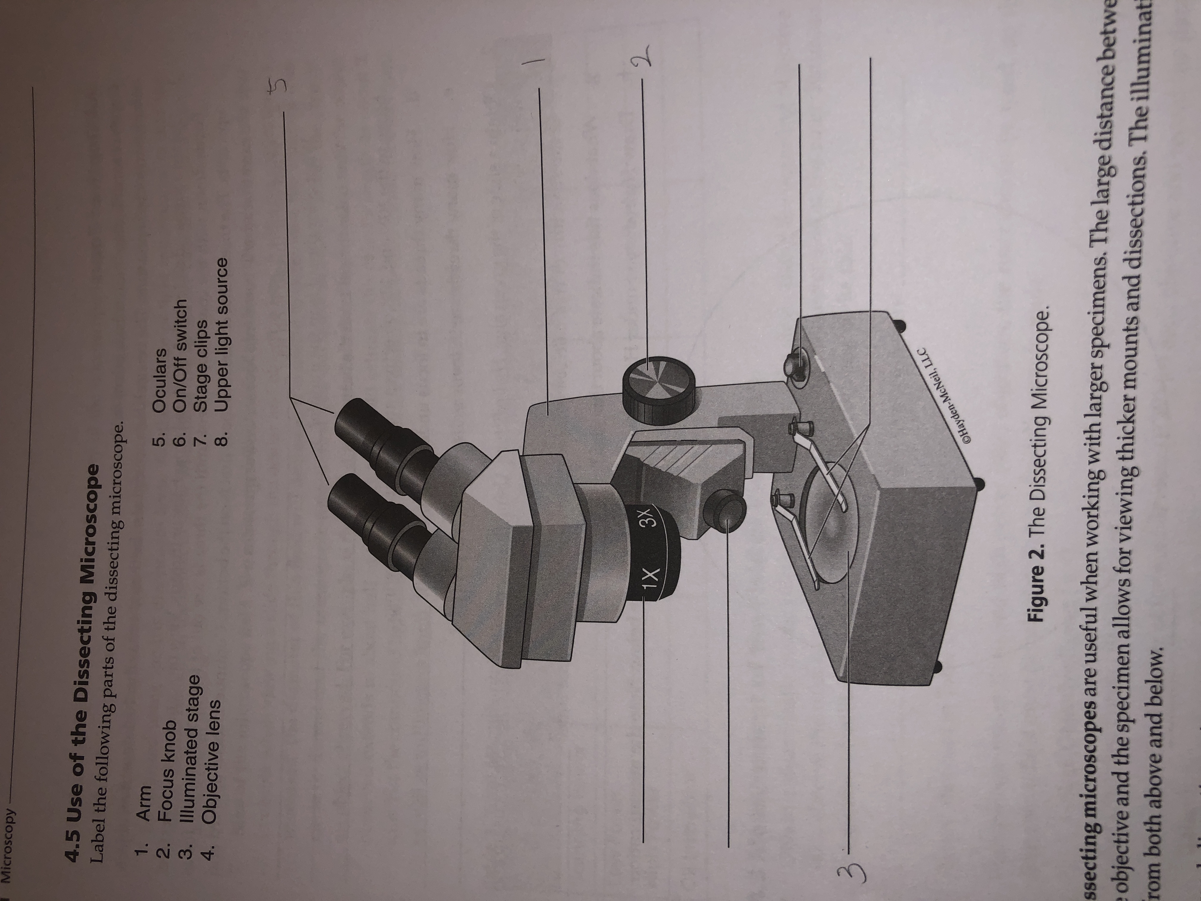
Answered: Microscopy 4.5 Use of the Dissecting… | bartleby
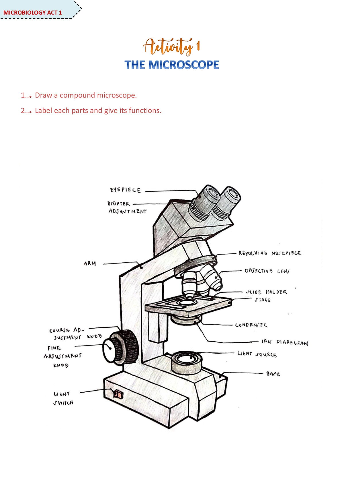
Microscope Activity - MICROBIOLOGY - 1... Draw a compound ...
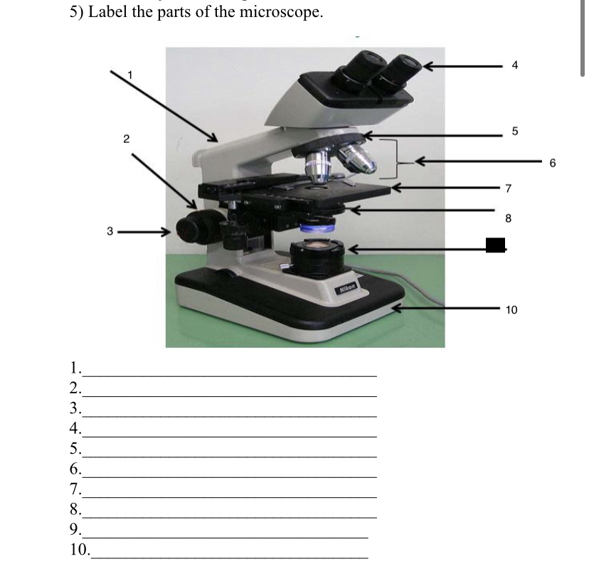
Answered: 5) Label the parts of the microscope. 1… | bartleby
Compound Microscope Parts, Functions, and Labeled Diagram ...

Parts of a Microscope | Labeling activities, Science ...

Microscope Labeling Diagram | Quizlet
The Parts of a Microscope (Labeled) Printable Printable (6th ...
Parts of the Microscope Labeling Activity!

Microscope Terms Glossary | Earth science lessons, Biology ...
Parts of Stereo Microscope (Dissecting microscope) – labeled ...

Parts of a microscope with functions and labeled diagram
Post a Comment for "38 microscope images with labels"