38 diagram of a cell with labels
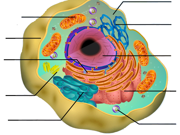
Learn the parts of a cell with diagrams and cell quizzes Labeled cell diagram For this exercise we'll start with an image of a cell diagram ready labeled. Study this and make sure that you're clear about which structure is found where. Cell diagram unlabeled It's time to label the cell yourself! Human Cell Diagram, Parts, Pictures, Structure and Functions Diagram of the human cell illustrating the different parts of the cell. Cell Membrane. The cell membrane is the outer coating of the cell and contains the cytoplasm, substances within it and the organelle. It is a double-layered membrane composed of proteins and lipids. The lipid molecules on the outer and inner part (lipid bilayer) allow it to ...
Label Cell Parts | Plant & Animal Cell Activity | StoryboardThat Create a cell diagram with each part of plant and animal cells labeled. Include descriptions of what each organelle does. Click "Start Assignment". Find diagrams of a plant and an animal cell in the Science tab. Using arrows and Textables, label each part of the cell and describe its function.

Diagram of a cell with labels
03 Label the Cell Diagram | Quizlet Cell Biology 03 Label the Cell STUDY Learn Flashcards Write Spell Test PLAY Match Gravity Created by muskopf1TEACHER Terms in this set (14) Nucleus Control center of the cell Nucleolus Ribosome synthesis Rough Endoplasmic Reticulum Protein transport Smooth Endoplasmic Reticulum Lipid synthesis Mitochondrion Cellular Respiratoin Golgi Apparatus Diagram Of Plant And Animal Cells To Label Teaching Resources | TpT Google Drive™ folder. This worksheet includes both an extensive animal and plant cell diagram to label. The animal cell includes 17 organelles, and the plant cell includes 20 organelles for students to label and color. There is also a 4 page graphic organizer (chart) that includes a drawing of each of the organelles in alphabetical order. Printable Animal Cell Diagram - Labeled, Unlabeled, and Blank - Tim's ... A printable diagram of an animal cell. This PDF includes the color version, black and white version, and the labeled and unlabeled diagrams for students to complete. 6 pages total.
Diagram of a cell with labels. How to draw an animal cell - labeled science diagram - YouTube Download a free printable outline of this video and draw along with us: you for watching. Please ... How to draw a nerve cell - labeled science diagrams - YouTube Download a free printable outline of this video and draw along with us: you for watching. Please su... Plant Cells: Labelled Diagram, Definitions, and Structure Plant Cells: Labelled Diagram, Definitions, and Structure Structure of Plant Cells Cell Wall Plant cells are eukaryotic cells, but unlike animal cells which have a cell membrane, plant cells have cell walls. Plants have a rigid cell wall that surrounds the plasma membrane. Interactive Bacteria Cell Model - CELLS alive Periplasmic Space: This cellular compartment is found only in those bacteria that have both an outer membrane and plasma membrane (e.g. Gram negative bacteria).In the space are enzymes and other proteins that help digest and move nutrients into the cell. Cell Wall: Composed of peptidoglycan (polysaccharides + protein), the cell wall maintains the overall shape of a …
Chord diagram – from Data to Viz A chord diagram represents flows or connections between several entities (called nodes).Each entity is represented by a fragment on the outer part of the circular layout.Then, arcs are drawn between each entities. The size of the arc is proportional to the importance of the flow. Here is an example displaying the number of people migrating from one country to another. Labeled Plant Cell With Diagrams | Science Trends The parts of a plant cell include the cell wall, the cell membrane, the cytoskeleton or cytoplasm, the nucleus, the Golgi body, the mitochondria, the peroxisome's, the vacuoles, ribosomes, and the endoplasmic reticulum. Parts Of A Plant Cell The Cell Wall Let's start from the outside and work our way inwards. A Labeled Diagram of the Animal Cell and its Organelles A Labeled Diagram of the Animal Cell and its Organelles There are two types of cells - Prokaryotic and Eucaryotic. Eukaryotic cells are larger, more complex, and have evolved more recently than prokaryotes. Where, prokaryotes are just bacteria and archaea, eukaryotes are literally everything else. animal and plant cell diagram to label - TeachersPayTeachers 12. $2.00. PDF. Three versions of the plant cell worksheet and three versions of the animal cell worksheet allow students of different grade levels and/or skill levels to label and review the parts of each cell type. Can be used as homework or a quiz. Answer key is provided and can be projected and used during less.
Bacteria in Microbiology - shapes, structure and diagram Bacterial spores. Bacterial endospores layers. Bacteria cells are the smallest living cells that are known; even though viruses are smaller than bacteria, viruses are not living cells. There are different types of bacteria with various sizes, shapes, and structures. The bacteria shapes, structure, and labeled diagrams are discussed below. Cells Diagram | Science Illustration Solutions - Edrawsoft Edraw software offers you lots of symbols used in cells diagram like cell structure, paramecium, squamous cell, cell division, bacteria, cell membrane, eggs, sperm, zygote, an animal cell, SARS, tobacco mosaic, adenovirus, coliphage, herpesvirus, AIDS, pollen, plant cell model, onion tissue, etc. Cells Diagram Examples Examine the diagram of a cell. Which accurately labels the lysosome? W ... Explanation: Lysosomes are heterogeneous structures present in animal cells which are bound by single membranes. They are of varying shape and size and contains hydrolytic enzymes inside it. Lysosomal membrane has H⁺ ATPase which pumps H⁺ into the membrane through ATP hydrolysis. This pumping of H⁺ makes the internal pH of lysosome acidic. Coordination Chemistry III: Tanabe-Sugano Diagrams and Charge … Symmetry Labels for Configurations Free ion termssplit into statesin the ligand field, according to symmetry: The state labels also indicate the degeneracy of the electron configuration: d7Tanabe-Sugano Diagram E / B ∆o/ B 4F 2G 2Eg 2T1g 2A1g 2T2g 4P 4A 2g 4T 1g (4P) 4T 2g 4T 1g (4F) Complexes with d4-d7 electron counts are special •at small values of ∆o/B the diagram looks …
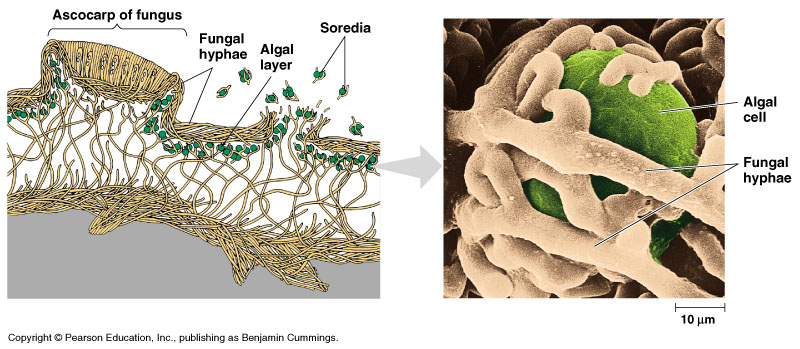
Fungi
Cell Organelles- Definition, Structure, Functions, Diagram In a plant cell, the cell wall is made up of cellulose, hemicellulose, and proteins while in a fungal cell, it is composed of chitin. A cell wall is multilayered with a middle lamina, a primary cell wall, and a secondary cell wall. The middle lamina contains polysaccharides that provide adhesion and allow binding of the cells to one another.

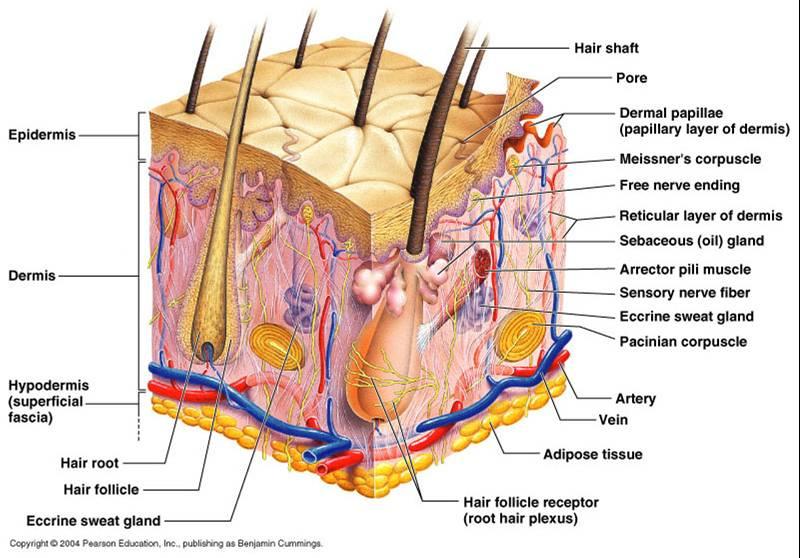
Label A Cell Diagram - Human Anatomy
Animal Cells: Labelled Diagram, Definitions, and Structure Plant cell are the basic unit of all plants. Plant cells, like animal cells, are eukaryotic, meaning they have a membrane-bound nucleus and organelles. Animal Cells Diagram Animal Cells Organelles and Functions Plant Cells Diagram Plant Cells Organelles and Functions Animals Cells Vs Plant Cells
Cell Diagram To Label - ClipArt Best
PDF Human Cell Diagram, Parts, Pictures, Structure and Functions Human Cell Diagram, Parts, Pictures, Structure and Functions 1 Human Cell Diagram, Parts, Pictures, Structure and Functions The cell is the basic functional in a human meaning that it is a self-contained and fully operational living entity.
Print Exercise 7 flashcards | Easy Notecards
diagram of a cell labeled Explain The Nucleus Of A Cell With A Neat Labeled Diagram - Science labeled cell nucleus diagram explain neat functions structure CH 467 PIGEON SKELETON | Dbios Charts ch pigeon skeleton zoology aves 3D Model Of Animal Cell - YouTube

Attack of The Silverfish!: Images of the stinging cells of Cnidaria
A Well-labelled Diagram Of Animal Cell With Explanation Well-Labelled Diagram of Animal Cell The Cell Organelles are membrane-bound, present within the cells. There are various organelles present within the cell and are classified into three categories based on the presence or absence of membrane. Listed below are the Cell Organelles of an animal cell along with their functions.
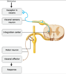
Print A&P Chapter 14 The Autonomic Nervous System flashcards | Easy Notecards
Drawing & Labeling a Diagram of a Electrochemical Cell - Study.com Lesson Summary. An electrochemical cell consists of two half-cells connected by a salt bridge.Each half-cell consist of a metal strip in a solution of that metal. Oxidation occurs in one half-cell ...
Cell Diagram To Label - ClipArt Best
Cell Diagrams - The Biology Corner Open Google Draw and import the diagram. Then use "insert" to create text boxes where students can fill in the labels. Don't forget when assigning this to students on Google classroom to make a copy for each student. You can leave documents in an uneditable form and students can use an addon like "Kami" to annotate the document.
Parts and Function of Digestive System for Med School & Nursing Students - NCLEX Quiz
How to Create Venn Diagram in Excel – Free Template Download Step #6 – Create the Diagram Labels: ... Step #4: Outline the x- and y-axis values for the Venn diagram circles. In a blank cell near the table with your data, map out the x- and y-axis coordinates which will be used as the centers of the circles. The following values are constants that will determine the position of Venn diagram circles on the chart plot, giving you full control …
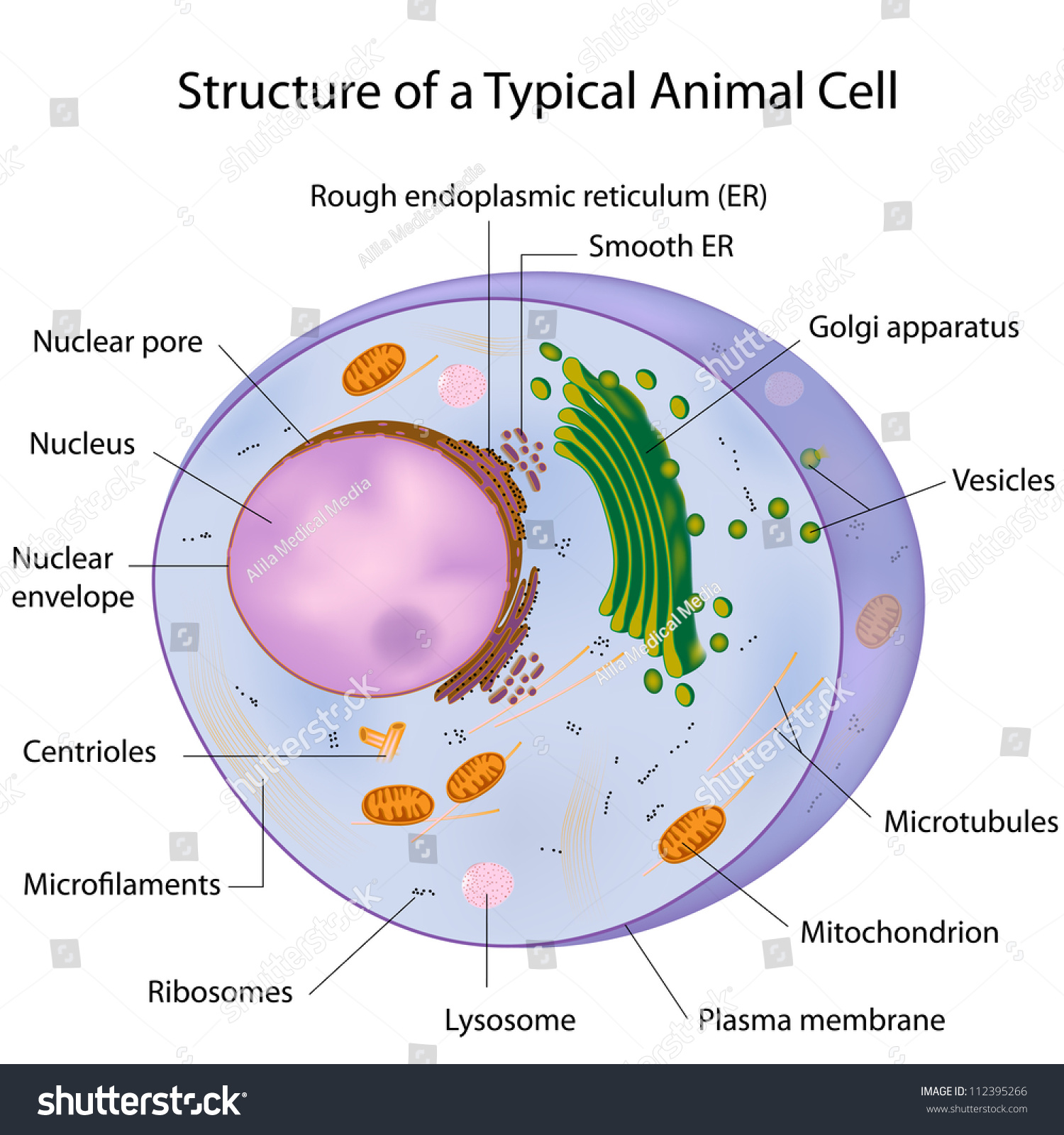
Typical Cell Labeled Stock Illustration 112395266 - Shutterstock
Cell: Structure and Functions (With Diagram) - Biology Discussion Eukaryotic Cells: 1. Eukaryotes are sophisticated cells with a well defined nucleus and cell organelles. 2. The cells are comparatively larger in size (10-100 μm). 3. Unicellular to multicellular in nature and evolved ~1 billion years ago. 4. The cell membrane is semipermeable and flexible. 5. These cells reproduce both asexually and sexually.

label diagram of cell
Cell Diagram | Free Cell Diagram Templates - Edrawsoft Cell Diagram Template Download Template: Get EdrawMax Now! Free Download Popular Latest Flowchart Process Flowchart Workflow BPMN Cross-Functional Flowchart Data Flow Diagram EPC Fault Tree IDEF Diagram Org Chart Basic Org Chart Photo Org Chart Creative Org Chart Family Tree Genogram Network Rack Diagram Network Topology CCTV Network LDAP

A typical cell, labeled diagram. | Alila Medical Images
› graph › chordChord diagram – from Data to Viz A chord diagram represents flows or connections between several entities (called nodes). Each entity is represented by a fragment on the outer part of the circular layout . Then, arcs are drawn between each entities.
Quia - Meiosis Illustration Identification
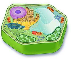
A Labeled Diagram of the Plant Cell and Functions of its Organelles A Labeled Diagram of the Plant Cell and Functions of its Organelles We are aware that all life stems from a single cell, and that the cell is the most basic unit of all living organisms. The cell being the smallest unit of life, is akin to a tiny room which houses several organs. Here, let's study the plant cell in detail...
Plant cell - SignWiki
Animal Cell Diagram with Label and Explanation: Cell Structure, Functions Below is the diagram of the animal cell which shows the organelles present in it. The cell is covered with cytoplasm which consists of cell organelles in it. The nucleus is covered with a rough Endoplasmic Reticulum and other organelles each designed for a specific purpose.

My Classroom
Plant Cell Diagram | Science Trends A plant cell diagram, like the one above, shows each part of the plant cell including the chloroplast, cell wall, plasma membrane, nucleus, mitochondria, ribosomes, etc.A plant cell diagram is a great way to learn the different components of the cell for your upcoming exam. Plants are able to do something animals can't: photosynthesize.Plant cells are able to do this because plant cells have ...
CELL - Labelled diagram
Labeling a Cell Diagram | Quizlet Cell Wall This gives shape and support to the plant cell. It surrounds the cell and protects the other parts of the cell. Chloroplasts This is where the plant cell's chlorophyll is stored. This is what the plant uses to make its own food (photosynthesis). This is also what makes plant cells have a green-like color. Plant cells Are circular in shape
Cell functions - Teaching resources
› ~lawm › extraCoordination Chemistry III: Tanabe-Sugano Diagrams and Charge ... d7Tanabe-Sugano Diagram E / B ∆o/ B 4F 2G 2Eg 2T1g 2A1g 2T2g 4P 4A2g 4T1g (4P) 4T2g 4T1g (4F) small ∆o High Spin large ∆o Low Spin Complexes with d4-d7 electron counts are special •at small values of ∆o/B the diagram looks similar to the d2diagram •at larger values of ∆o/B, there is a break in the diagram leading to a new ground ...
Chapter- 9 Cellular Respiration and Fermentation - Part A Flashcards | Easy Notecards
help.devexpress.comOnline Documentation - Developer Express Inc. Buy Support Center Documentation Blogs Training Demos Free Trial Log In

label diagram of cell
Printable Animal Cell Diagram - Labeled, Unlabeled, and Blank - Tim's ... A printable diagram of an animal cell. This PDF includes the color version, black and white version, and the labeled and unlabeled diagrams for students to complete. 6 pages total.
Post a Comment for "38 diagram of a cell with labels"